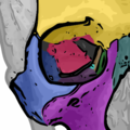
Orbital_bones.png (350 × 350 pixels, file size: 104 KB, MIME type: image/png)
File history
Click on a date/time to view the file as it appeared at that time.
| Date/Time | Thumbnail | Dimensions | User | Comment | |
|---|---|---|---|---|---|
| current | 20:19, 17 November 2007 |  | 350 × 350 (104 KB) | ToNToNi | {{Information |Description={{en|This picture, adapted from Gray's Anatomy [[:en::Image:Gray190.png|fig. 190]], highlights each of the 7 bones that form the orbit of the eye. It was made using Inkscape and Photoshop CS, by {{User:je_at_uwo/sig}} Yellow: f |
File usage
The following 19 pages use this file:
- Ethmoid bone
- Greater wing of sphenoid bone
- Lacrimal bone
- Nasolacrimal canal
- Optic canal
- Orbit (anatomy)
- Orbital lamina of ethmoid bone
- Orbital part of frontal bone
- Orbital process of palatine bone
- Palatine bone
- Rhinoplasty
- Sphenoid bone
- User:Je at uwo
- User:Þjarkur/Rhinoplasty
- User talk:Je at uwo
- Wikipedia:Featured picture candidates/January-2015
- Wikipedia:Featured picture candidates/Orbital bones
- Wikipedia:WikiProject Anatomy/Resources
- Wikipedia talk:WikiProject Anatomy/Archive 9
Global file usage
The following other wikis use this file:
- Usage on ar.wikipedia.org
- Usage on bn.wikipedia.org
- Usage on bs.wikipedia.org
- Usage on ca.wikipedia.org
- Usage on cs.wikipedia.org
- Usage on da.wikipedia.org
- Usage on de.wikipedia.org
- Usage on es.wikipedia.org
- Usage on eu.wikipedia.org
- Usage on fr.wikipedia.org
- Usage on gl.wikipedia.org
- Usage on he.wikipedia.org
- Usage on hi.wikipedia.org
- Usage on hu.wikipedia.org
- Usage on hy.wikipedia.org
- Usage on it.wikipedia.org
- Usage on ka.wikipedia.org
- Usage on ko.wikipedia.org
- Usage on ku.wikipedia.org
- Usage on lt.wikipedia.org
- Usage on mk.wikipedia.org
View more global usage of this file.


