Abstract
An international group of clinicians working in the field of dysmorphology has initiated the standardization of terms used to describe human morphology. The goals are to standardize these terms and reach consensus regarding their definitions. In this way, we will increase the utility of descriptions of the human phenotype and facilitate reliable comparisons of findings among patients. Discussions with other workers in dysmorphology and related fields, such as developmental biology and molecular genetics, will become more precise. Here we describe the general background of the project and the various issues we have tried to take into account in defining the terms.
Keywords: nomenclature, definitions, morphology, dysmorphology, birth defects, malformations, minor anomalies, common variants
Introduction
This issue of the Journal contains six articles that describe the initial results of a project intended to develop accurate and clear definitions of terms for the craniofacies in general, the major components of the face, and the hands and feet (Allanson et al., 2009; Biesecker et al, 2009; Carey et al., 2009; Hall et al., 2009; Hennekam et al., 2009; Hunter et al., 2009). These articles are the result of a significant amount of planning, organization, negotiation, review, and writing, while, at the same time, they are but a start.
Dysmorphology evolved from a small nucleus of clinicians in the 1950s into a recognized and widely practiced discipline, and more recently has incorporated translational research into developmental biology, molecular genetics, and metabolic medicine. The terms that clinicians use to describe a body part have gradually evolved in a haphazard and uncoordinated manner, and have not been critically reviewed. Clinicians and researchers have always made comparisons among patients and syndromes, and in the last decade it has become increasingly possible and necessary to use clinical data for studies of etiology and pathogenesis, epidemiology, the isolation of causative gene mutations, and for evaluation of interventions. Therefore, we need to have uniform and internationally accepted terms to describe the human phenotype.
Organization of the Project
Several years ago, after many informal discussions, we brought together an international group of clinicians working in the field of dysmorphology with the goal of standardizing this nomenclature. The participants were chosen for their knowledge of morphology, syndromes, and skills in syndrome delineation. Because we anticipated that the group would need to gather several times and we recognized financial constraints, we restricted the group to 34 individuals and had to accept that many excellent colleagues could not be invited to join the project.
Our original goal was to review, revise if necessary, and delineate definitions for all of the terms used in the London Dysmorphology Databases (n=683). However, recognizing the enormity of the task, we chose to begin the project with the cranium, the face and its features, and the hands and feet – the parts of the body that are often used in dysmorphology to describe patients and delineate syndromes.
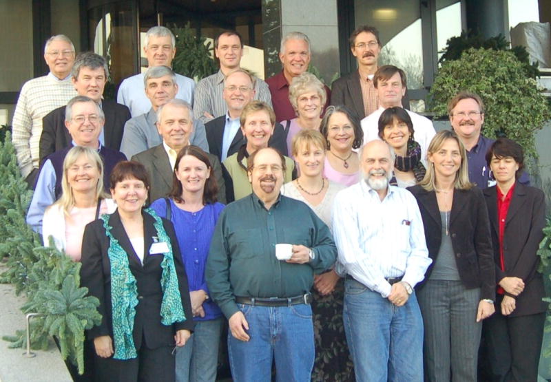
We set out to accomplish this work in six teams, each focused on a particular area of the human body. While much of our work has been carried out in small groups using electronic communication, we have met as a large group on two occasions: in Bethesda in December, 2005 (host: Leslie G Biesecker) and Rome in November, 2006 (hosts: Giovanni Neri and Fiorella Gurrieri) (Fig. 1). These face-to-face meetings were invaluable in expediting the process and allowing discussion of principles and initial points of disagreement, of which there were many.
Figure 1.

Nomenclature group members present in November 2006 in Rome (Suzanne Cassidy was also present but could not be depicted on this picture). From left to the right are visible (first row) Helga Toriello, Cynthia Curry, Julie McGaughran, M Michael Cohen Jr, Louise Wilson, John Carey, Fiorella Gurrieri, Valerie Cormier-Daire, (second row) Jaime Frias, Giovanni Neri, Judith Allanson, Judith Hall, Karen Temple, Alain Verloes, (third row) Michael Patton, Alasdair Hunter, Gene Hoyme, Helen Hughes, John M Graham Jr, (fourth row) Roger Stevenson, Leslie Biesecker, Koen Devriendt, Bryan Hall, and Raoul Hennekam.
Attributes of the Terms
For each feature we provide a preferred term and use a standard format to provide a definition and description of how to observe and measure (where possible) the feature. For many of the terms, both subjective and objective definitions of terms are provided. When an objective assessment can be made, this is always preferable to the subjective. A few terms have multiple alternative objective definitions. Typically, this reflects multiple sets of norms for a quantitative trait and, in such cases, each definition is equally valid.
Our aim is to formalize, unify, and standardize the approach to clinical assessment in the hope that it will become generally accepted and applied. For each term we have added relevant comments and synonyms, and indicated terms that should no longer be used. Most clinicians have at least a few terms of which they are inordinately fond. In spite of these personal biases, our goal is to make a universal language to describe morphology. We have avoided terms that indicate pathogenesis or an active process; we aspired to simply describe what can be observed.
We recognize that a number of terms in common usage may be considered pejorative in some or all cultures. We have created alternative descriptors in these cases, even if they are not in current common usage. The definitions are intended to be applied broadly, thus we avoided obscure terms, flowery language, etc. We have tried to apply a standard format that would be readily understandable by a medical student who has completed training in physical diagnosis.
In addition to standardized terms and definitions, we aimed to provide clear illustrations of each feature. We have used our joint collection of pictures and the extensive Robert J. Gorlin slide collection which is the series of digitalized slides that Prof Gorlin gathered during his long career available to one of us (RCMH). It is important that readers recognize that a given photograph may include multiple findings. Where possible, photographs have been cropped to avoid showing other signs and to emphasize one feature. We tried to show variable expression of the features, both mild and severe.
The terms are intended for clinical evaluation of the individual using observation, palpation, or simple morphometric techniques (such as measuring of inner canthal distance). We provide basic general principles for measuring each of the body parts, and indicate in the definitions if a reliable measurement is possible and if norms are available. However, extensive description of morphometric techniques is outside the scope of this effort and readers should consult authoritative sources for this information (Farkas L Anthropometry of the Head and Face in Medicine. Elsevier, 1981; and Hall JG, Allanson JE, Gripp KW, Slavotinek AM. Handbook of Physical Measurements, 2nd Edition, OUP, 2007). We have referenced the most important standards for each of the body parts. The present project was not intended to generate new normative data, novel objective assessment techniques, or novel morphologic features. We acknowledge a tremendous lack of data on ethnic groups other than Caucasian, and we encourage efforts to address this deficiency.
The use of radiographs as part of morphologic assessments (such as a hand radiograph to distinguish a broad thumb from a bifid thumb) was controversial in the group. Although we decided not to use radiographic findings in the definitions and only use data obtained through surface examination, this issue will need to be re-evaluated.
We provide only limited and general anatomical background and have avoided more substantive explanations and reviews of what is known (or hypothesized) about the mechanism of the genesis of the physical findings. This is for several reasons. First, other excellent texts provide thorough discussions of this topic (Sadler TW, Langman J Langman's Medical Embryology 8th Ed, Lippincott Williams & Wilkins, 2000; Moore KL, Dalley AF Clinical Oriented Anatomy, 5th Ed, Lippincott Williams & Wilkins, 2005). Second, our understanding of the genesis of an anatomical finding is likely to change over time, and we did not want the definitions to change should this occur. Third, the current debate about the proposed genesis of many of the findings could detract from the intended goals of this work.
We emphasize that the terms deliberately avoid any commentary about normal or abnormal status, even when this is obvious (such as a proboscis). This is because many morphological findings are commonly observed as isolated features in the normal population. There can also be a remarkable difference in frequency of a finding among various populations. The objectively determined findings are by definition abnormal in a normative, statistical sense. However, even in these cases, the individual should simply be considered to have the feature, and no judgment of normal or abnormal status is made.
We have elected not to provide differential diagnoses for the features as such lists could be very long or incomplete. Syndromes are typically diagnosed by combinations of features, and therefore differential diagnoses of single features are of limited utility. Readers should consult databases of medical dysmorphology for this information (London Dysmorphology Databases, version 1.0.14, 2008; POSSUM web version 2008).
Bundling is a word we have used to describe a term that represents two or more component findings. The term “large nose” can serve as an example of a bundled term as the term comprises several distinct features: prominent nose; wide nasal ridge; prominent nasal tip; and broad nasal base. We have eliminated most bundled terms as they often include presumptions of pathogenesis or association, which may or may not be correct. Bundling is also problematic because it can obscure component abnormalities and if individuals are described using both the single bundled term and the component terms, it can lead to confusion. A number of terms have remained bundled, as we felt that the utility of the bundled term outweighed the potential pitfalls of bundling. If a term is considered to be bundled, this is mentioned in the definition and the individual components are indicated in the description.
The Future of the Terminology Project
While the project thus far has taken more than five years, there is a long road ahead. The present series of papers should be considered a starting point, and we enthusiastically solicit input from colleagues within and beyond the field of dysmorphology on some of the difficult issues described here to correct, edit, modify, or extend the definitions. We acknowledge that we are a self-selected special interest group and do not claim to represent the international dysmorphology community. We want the definitions to be available and useful to a large community of clinicians and researchers. Thus, we plan to distribute them to other expert groups including dentists, ophthalmologists, dermatologists and other non-geneticists for comments in their area of expertise. Our intent is to use this input to refine the terms and definitions, add the body parts that we have not yet defined, and publish the complete group of terms needed to describe the human phenotype as a monograph. With time we hope to make them available in other languages.
We are presently working on adapting the working groups that established the definitions presented here into a permanent international nomenclature committee, such as the ones that exist in cytogenetics (International Standard of Cytogenetic Nomenclature) and molecular genetics (The Human Genome Variation Society nomenclature recommendations). Such a committee could periodically review the definitions and change them where necessary. Edward Sapir (1884-1939) is famous for his axiom “Language structures thought”. We hope our joint effort will help to structure the language we use in dysmorphology.
Table. Nomenclature Subgroups and their members.
(the chair of each group is in italics)
| Head and Face: Judith E. Allanson Chris Cunniff Gene Hoyme Julie McGaughran Max Muenke Giovanni Neri |
| Hands and Feet: Leslie G. Biesecker Jon M. Aase Carol Clericuzio Fiorella Gurrieri Karen Temple Helga Toriello |
| Mouth: John C. Carey M. Michael Cohen Jr Cynthia Curry Koen Devriendt Lewis Holmes Alain Verloes |
| Ear: Alasdair G. W. Hunter Jaime Frias Gabrielle Gillessen-Kaesbach Ken Jones Helen Hughes Louise Wilson |
| Periorbital structures: Bryan D. Hall John M. Graham Jr Suzanne B. Cassidy John M. Opitz |
| Nose and Philtrum: Raoul C. M. Hennekam Valerie Cormier-Daire Judith G. Hall Karoly Méhes Michael Patton Roger Stevenson |
Acknowledgments
We are grateful for financial support from the Birth Defects Foundation – Newlife (UK), Institute of Child Health (UK), National Foundation of the March of Dimes (USA), National Human Genome Research Institute (USA), Catholic University of Rome (Italy), and John Wiley and Sons publishing. In the initial phase of the project Drs Heval Ozgen and Jan Maarten Cobben (Amsterdam) provided invaluable input.
References
- Allanson JE, Cunniff C, Hoyme HE, McGaughran J, Muenke M, Neri G. Elements of morphology: head and face. Am J Med Genet. 2009 doi: 10.1002/ajmg.a.32612. [DOI] [PMC free article] [PubMed] [Google Scholar]
- Biesecker LG, Aase JM, Clericuzio C, Gurrieri F, Temple K, Toriello H. Elements of morphology: hands and feet. Am J Med Genet. 2009 doi: 10.1002/ajmg.a.32596. [DOI] [PMC free article] [PubMed] [Google Scholar]
- Carey JC, Cohen MM, Jr, Curry C, Devriendt K, Holmes L, Verloes A. Elements of morphology: oral region. Am J Med Genet. 2009 doi: 10.1002/ajmg.a.32602. [DOI] [PubMed] [Google Scholar]
- Hall BD, Graham JM, Jr, Cassidy SB, Opitz JM. Elements of morphology: periorbital area. Am J Med Genet. 2009 doi: 10.1002/ajmg.a.32597. [DOI] [PubMed] [Google Scholar]
- Hennekam RCM, Cormier-Daire V, Hall J, Méhes K, Patton M, Stevenson R. Elements of morphology: nose and philtrum. Am J Med Genet. 2009 doi: 10.1002/ajmg.a.32600. [DOI] [PubMed] [Google Scholar]
- Hunter A, Frias J, Gillessen-Kaesbach G, Hughes H, Jones K, Wilson L. Elements of morphology: Ear. Am J Med Genet. 2009 doi: 10.1002/ajmg.a.32599. [DOI] [PubMed] [Google Scholar]
