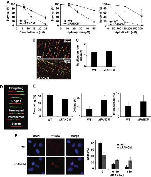
Figure 1.
FANCM-deficient cells are hypersensitive to agents interfering with replication, display altered global DNA replication, and accumulate spontaneous DNA damage. (A) Cellular sensitivity of WT and ΔFANCM cells to camptothecin (CPT), hydroxyurea (HU), and aphidicolin (APH) as measured by MTS survival assay. Mean values of three independent experiments are shown ±s.e.m. (B) Representative images of actual fibres from WT and ΔFANCM cells. (C) Replication fork velocity in the WT DT40 and ΔFANCM cells used in this study. A minimum of 200 tracts were measured per experiment. Mean values for three independent experiments are shown ±s.e.m. (D) Five classes of replication structures and (E) the relative frequency of occurrence of these different classes. A minimum of 200 fibres were scored per experiment. Mean values of three independent experiments are shown ±s.e.m. (F) γ-H2AX foci in untreated WT and ΔFANCM cells. Left panel shows representative images of γ-H2AX foci. Right panel shows quantification of percentage cells exhibiting γ-H2AX foci out of more than 200 cells per experiment. Bars represent mean values from three independent experiments ±s.e.m.
