Figure 3.
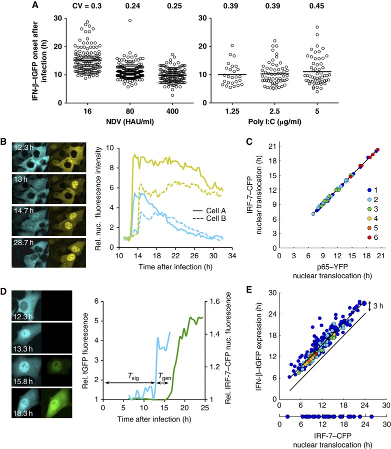
Temporal variability in cellular IFN-β induction. (A) IFN-β expression onset in single cells. Variability of response timing is virus-independent. IFN-β–tGFP reporter cells infected for 1 h with indicated concentrations of NDV or transfected with poly I:C at given concentrations were subjected to time-lapse microscopy (15 min picture intervals). Distribution of tGFP expression onset over time (scatter plot, n=456 (NDV), n=140 (poly I:C)) and CVs are shown. (B) Synchronous activation of NF-κB and IRF-7. NIH3T3 cell clone stably expressing the fusion proteins IRF-7–CFP and NF-κB/p65–YFP were infected with 80 HAU/ml NDV for 1 h and subjected to time-lapse microscopy. Fluorescence pictures for CFP and YFP were taken every 20 min. Subcellular localization of IRF-7–CFP (left column) and p65–YFP (right column) at indicated time after infection. The diagram shows relative nuclear fluorescence for IRF-7–CFP and p65–YFP from sister cells. (C) Synchronicity is independent of response time. IRF-7–CFP and p65–YFP initial nuclear translocation were determined in individual cells and plotted against each other (n=65). Coloured dots indicate the frequency of data points. (D) Expression delay of an individual cell. NIH3T3 cell clone stably expressing IRF-7–CFP together with IFN-β–tGFP were infected with 80 HAU/ml NDV. Fluorescence pictures for CFP and GFP were taken every 20 min. Subcellular localization of IRF-7–CFP (left column) and IFN-β–tGFP expression (right column) at indicated time after infection. Graphs show relative IRF-7–CFP nuclear fluorescence and tGFP intensity. Tsig: time interval between infection and IRF-7–CFP nuclear translocation. Tgen: time interval between IRF-7–CFP nuclear translocation and onset of IFN-β–tGFP gene expression. (E) Response variation at distinct stages of IFN induction. The starting times for IRF-7–CFP nuclear translocation were plotted against the times of IFN-β–tGFP expression for individual cells (n=315). Source data is available for this figure in the Supplementary Information.
