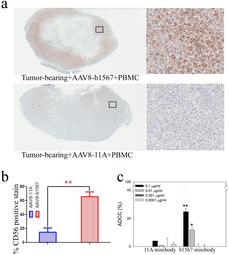
Figure 4. ADCC activity of h1567 minibody in a xenograft human PBMC-SCID/BEIGE mouse model.
(a) Immunohistochemical staining of a representative tumor section with mAb directed against human NK cell surface marker CD56. The immunostaining shows highly positive CD56 tumor-infiltrating human NK cells (brown stain) in tumor from the SCID/BEIGE mice treated with AAV8-h1567 and hPBMCs (upper panel). Negative CD56 staining was seen in the tumor treated with control vector AAV8-11A plus hPBMCs (lower panel). Images are shown from whole tumor cut sections (left panels) and tumor sections at 20× magnifications (right panels). (b) The percentage of immunohistochemically detected tumor-infiltrating natural killer cells was plotted. A significantly higher percentage of tumor-infiltrating human CD56-positive cells were detected in the AAV8-h1567-treated mice group. **, p<0.01. (c) NK cell-mediated cytotoxicity was observed in a dose-dependent manner. Minibody concentrations from 0.0001 to 0.1 ug/ml were tested at an E:T ratio of 2∶1. The average and error bars (mean + SD) shown were calculated from triplicate wells of one experiment. The figures shown are representative of three independent experiments. *P<0.05, **P<0.01 when comparing h1567 minibody-treated and 11A control minibody-treated group. All data is shown as the mean ± SD.
