Abstract
Regulatory T cells (Tregs) are key mediators of immune tolerance and feature prominently in cancer. Depletion of CD25+ FoxP3+ Tregs in vivo may promote T cell cancer immunosurveillance, but no strategy to do so in humans while preserving immunity and preventing autoimmunity has been validated. We evaluated the Food and Drug Administration–approved CD25-blocking monoclonal antibody daclizumab with regard to human Treg survival and function. In vitro, daclizumab did not mediate antibody-dependent or complement-mediated cytotoxicity but rather resulted in the down-regulation of FoxP3 selectively among CD25high CD45RAneg Tregs. Moreover, daclizumab-treated CD45RAneg Tregs lost suppressive function and regained the ability to produce interferon-γ, consistent with reprogramming. To understand the impact of daclizumab on Tregs in vivo, we performed a clinical trial of daclizumab in combination with an experimental cancer vaccine in patients with metastatic breast cancer. Daclizumab administration led to a marked and prolonged decrease in Tregs in patients. Robust CD8 and CD4 T cell priming and boosting to all vaccine antigens were observed in the absence of autoimmunity. We conclude that CD25 blockade depletes and selectively reprograms Tregs in concert with active immune therapy in cancer patients. These results suggest a mechanism to target cancer-associated Tregs while avoiding autoimmunity.
INTRODUCTION
Regulatory T cells (Tregs) protect against autoimmunity, but in cancer, Tregs infiltrate even the earliest neoplastic lesions and undermine anti-tumor effector T cells (1–4). Treg development and homeostasis are critically dependent on interleukin-2 (IL-2) (5–9), and most Tregs express high levels of CD25, the cell surface α chain of the IL-2 receptor (3). In tumor-bearing mice, CD25 monoclonal antibody (mAb) can be used to deplete CD25+ Tregs in vivo and enhance tumor immunity and immunotherapy (10–12); however, the clinical translation of this approach may pose a risk of autoimmunity (13, 14) or interfere with effector T cells (12) or myeloid dendritic cells (15, 16) that transiently express CD25 upon activation.
We reconsidered these concerns in light of landmark studies demonstrating that IL-2 is largely dispensable for effector T cell responses (17) and also because recent data suggest that IL-2 deprivation can regulate plasticity in Treg gene expression (18, 19). Daclizumab is a Food and Drug Administration (FDA)–approved mAb with a prolonged half-life that blocks IL-2 binding to CD25 (20). We and others hypothesized that long-term CD25 blockade accomplished with daclizumab would negatively impact human Tregs but spare or enhance effector T cell function (21, 22). Here, we tested this hypothesis both in vitro with human cells and in vivo in a clinical trial.
RESULTS
Effects of CD25 blockade in vitro
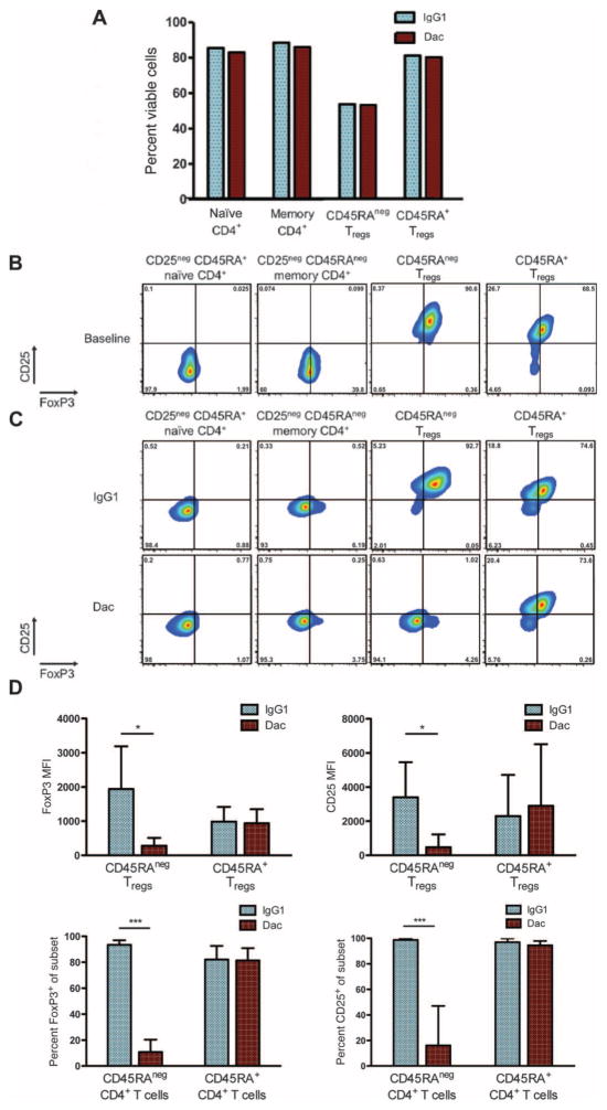
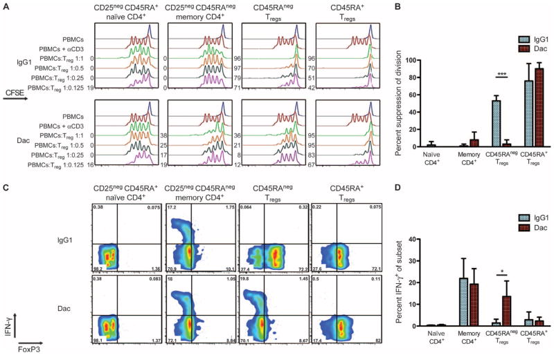
We first demonstrated in vitro that daclizumab does not elicit antibody-dependent cellular cytotoxicity or complement-mediated cytotoxicity, as previously noted (23), and also showed that binding of daclizumab is not acutely toxic for CD25-expressing cells (fig. S1). We then purified Treg populations from normal donors, examining CD4+ CD25high CD45RA+ T cells and CD4+ CD25high CD45RAneg T cells separately, because these Treg populations are known to differ in their stability, survival, epigenetic status of the Treg-specific demethylated region, and ability to produce IL-10 (24–26). For each Treg population, purified cells expressed FoxP3, the master regulatory transcription factor for Tregs (3, 4), whereas naïve and memory CD4 T cells isolated from the same donors as a control were FoxP3low or FoxP3neg. Upon incubation with IL-2 and either daclizumab or human immunoglobulin G1 (IgG1) in vitro, we found no difference in cell number or viability of any T cell subset when comparing daclizumab incubation versus control incubation (Fig. 1A). For CD45RAneg Tregs, expression of both FoxP3 and CD25 was markedly reduced in the presence of daclizumab compared to IgG1 in terms of both mean fluorescence intensity and percent positivity (Fig. 1, B to D). Concomitant with reduced FoxP3 and CD25 expression, daclizumab-incubated CD45RAneg Tregs were also less capable of suppressing anti-CD3 mAb-stimulated polyclonal allogeneic CD8 T cells compared to control-incubated Tregs (Fig. 2, A and B). Moreover, CD45RAneg Tregs were able to secrete interferon-γ (IFN-γ) in response to mitogens after daclizumab incubation, but not after incubation with IgG1 (Fig. 2, C and D). In contrast, CD45RA+ Tregs were unaffected by daclizumab with regard to FoxP3 and CD25 expression, suppressive function, and cytokine production (Figs. 1, B to D, and 2). These results suggest that CD25 blockade in vitro leads to selective reprogramming (18) of the CD25high CD45RAneg Treg population.
Fig. 1.
CD25 blockade in vitro mediates change in expression of FoxP3 and CD25 by human CD45RAneg Tregs. Flow cytometric cell sorting was used to purify four CD4+ T cell populations from normal human peripheral blood: naïve (CD25neg CD45RA+), memory (CD25neg CD45RAneg), activated Tregs (CD25high CD45RAneg), and resting Tregs (CD25high CD45RA+). Equal numbers of each cell population were incubated in vitro for 4 days with IL-2 (20 U/ml) and either human IgG1 (10 μg/ml) or daclizumab (Dac, 10 μg/ml). (A) Cell viability at day 4. (B and C) FoxP3 and CD25 expression for each cell population at (B) baseline and (C) at day 4 for each experimental condition, shown for a representative experiment. The percentage of cells in each quadrant is shown. (D) Quantification of mean fluorescence index (MFI) and percent positivity for FoxP3 and CD25 expression on Tregs at day 4, comparing IgG1 to daclizumab conditions. Data are from four or five independent experiments and shown as means ± SD. *P < 0.05; ***P < 0.001.
Fig. 2.
CD25 blockade in vitro mediates functional reprogramming of FoxP3 and CD25 human CD45RAneg Tregs. Equal numbers of each cell population were incubated in vitro with IgG1 or daclizumab (Dac), as in Fig. 1, and then tested for function. (A to D) Suppressive capability (A and B) and IFN-γ expression (C and D) upon phorbol 12-myristate 13-acetate (PMA) and ionomycin stimulation for each T cell population. Data shown in (A) and (C) are representative of three independent experiments. In (A), numbers shown on the left of each plot indicate percent suppression. In (C), the percentage of cells in each quadrant is shown. Summary data are shown in (B) and (D) as means ± SD. *P < 0.05; ***P < 0.001.
Clinical trial of daclizumab and cancer vaccination
To examine the impact of daclizumab on Tregs in vivo, we then performed a clinical trial for human leukocyte antigen–A2–positive (HLA-A2+) patients with metastatic breast cancer (table S1), a disease in which Tregs are especially prominent (27, 28). Patients received a single intravenous infusion of the CD25 mAb daclizumab (1 mg/kg) 1 week before receiving a series of subcutaneous injections of HLA-A2–binding peptides emulsified in adjuvant as part of a cancer vaccine (fig. S2). Three peptides were derived from hTERT (human telomerase reverse transcriptase), a fourth from survivin, and a fifth peptide (as a control) from pp65 of cytomegalovirus (CMV). Patients also received the seven-valent CRM197-containing pneumococcal conjugate vaccine (PCV) at the time of the first, third, and fifth peptide vaccine. Treatment was well tolerated (table S2), and there were no dose-limiting toxicities and no induction of autoimmune events. The best clinical response by RECIST (Response Evaluation Criteria in Solid Tumors) measurements was stable disease in 6 of 10 evaluable patients. Progression-free survival was 4.8 months [95% confidence interval (CI), 3.0 to 6.5 months], and median overall survival was 27.8 months (95% CI, 19.5 to 36.1 months) at a median follow-up of 22.3 months. Notably, at the 2-year benchmark, 65.5 ± 17.3% (rate ± SE) of patients were alive.
Effects of daclizumab on Tregs in vivo
At baseline in these patients, we observed that CD25high peripheral blood Tregs were heavily skewed toward a CD45RAneg CD45RO+ phenotype (fig. S3). After daclizumab infusion in vivo, we found statistically significant decreases in Tregs when measured as either CD25+ FoxP3+ CD4 T cells or total FoxP3+ CD4 T cells, whereas the overall CD4 T cell population did not change significantly over time (Fig. 3, A to C). This decrease in Tregs was rapid (occurring in less than 1 week), prolonged (lasting at least 7 weeks), and consistent (observed in 100% of patients). Results of linear random effects modeling demonstrated highly statistically significant decreases in the fraction of CD25+ FoxP3+ CD4 T cells at weeks 1, 2, 5, and 7, with mean reductions ranging from 56 to 77% (table S3). For total FoxP3+ CD4 T cells, significant decreases were observed at weeks 1 to 5, with mean reductions in the total Treg population of 52 to 64%. Results were nearly identical whether T cell subsets were calculated as fractions or absolute cell counts (Fig. 3, B and C).
Fig. 3.
CD25 blockade in vivo depletes systemic Tregs in cancer patients. Peripheral blood samples obtained from patients before and at various times after a single infusion of CD25 mAb daclizumab were analyzed by flow cytometry. (A) Representative data from one patient comparing baseline (before) to 5 weeks after daclizumab. (B and C) Relative changes in the fraction (B) or absolute counts (C) of various T cell subsets over time, shown as the means for all patients normalized to individual baseline values. Daclizumab was given on week 0. Red diamonds, total CD4+ T cells; blue squares, FoxP3+ CD4 T cells; purple triangles, CD25+ FoxP3+ CD4 T cells as identified by the nonblocked monitoring CD25 mAb 4E3; green circles, CD25+ FoxP3+ CD4 T cells as identified by the monitoring CD25 mAb 2A3, which is blocked by daclizumab. For total FoxP3+ CD4 T cells and CD25+ (4E3) FoxP3+ CD4 T cells, P < 0.001 at weeks 1, 2, and 5; for CD25+ (2A3) FoxP3+ CD4 T cells, P < 0.001 at weeks 1, 2, 5, and 7 (statistical details are provided in table S3). (D) Peptide vaccination of breast cancer patients without daclizumab administration does not alter Tregs. Peripheral blood T cell populations were analyzed as fractions before and after vaccination by flow cytometry in a cohort of patients (n = 7) with metastatic breast cancer on a previously described clinical trial (29). Nonblocked mAb 4E3 was used to evaluate CD25. Data are shown as the means of all patients normalized to individual baseline values with error bars representing SD. P > 0.05, comparing Treg fractions at the time of the second or third vaccine to baseline. Similar data were obtained if T cell populations were analyzed as absolute counts.
We ruled out that the monitoring CD25 mAb clone 4E3 is affected by the presence of daclizumab, and found further that daclizumab does not trigger endocytosis of the CD25 antigen (fig. S4). Using a second monitoring mAb that is fully blocked by daclizumab (clone 2A3) (fig. S4), we found a complete lack of 2A3 reactivity on FoxP3+ CD4 T cells after daclizumab (Fig. 3, B and C) that persisted for up to 7 weeks, indicating that daclizumab remains bound to CD25-expressing Tregs long-term, consistent with the drug’s 3-week serum half-life. The decreases in peripheral Tregs that we observed in this study were likely specific to daclizumab because vaccination of a comparable cohort of breast cancer patients with a nearly identical peptide formulation and schedule (29)—but without daclizumab—did not alter circulating Treg populations (Fig. 3D).
Recovery of peripheral blood Tregs was observed between 7 and 11 weeks after daclizumab infusion, parallel to the restoration of 2A3 reactivity (Fig. 3, B and C); however, for multiple patients analyzed at week 11 and beyond, Tregs never recovered to baseline. In these patients, CD25+ FoxP3+ CD4 T cells and total FoxP3+ CD4 T cells remained 20 to 60% below baseline levels (calculated as either fractions or absolute counts) (fig. S5). For six patients, the fractions and absolute counts of CD25+ FoxP3+ and total FoxP3+ Tregs reset within the range for healthy individuals of comparable age previously reported (28). Thus, daclizumab infusion also appears to reset Treg homeostasis to achieve a long-term reduction in the circulating burden of Tregs that is characteristic of cancer.
We also observed that CD25neg T cells (whether CD4 or CD8) are not affected by daclizumab administration (fig. S6). However, a small subset of FoxP3neg CD4 T cells and CD8 cells can also express CD25, and after daclizumab infusion, these cell populations decreased to about 40 to 65% of baseline (calculated as either fractions or absolute counts) before recovering by week 7 (fig. S6). In contrast, circulating CD56bright natural killer (NK) cells, which express little if any CD25, moderately increased after daclizumab (fig. S7), an effect previously reported in patients receiving daclizumab for multiple sclerosis, albeit via a repeated dosing schedule different than the single dose of daclizumab we used here (30, 31). Daclizumab had no statistically significant impact on CD56dull CD16bright NK cells (fig. S7).
Vaccine-induced immune responses after daclizumab
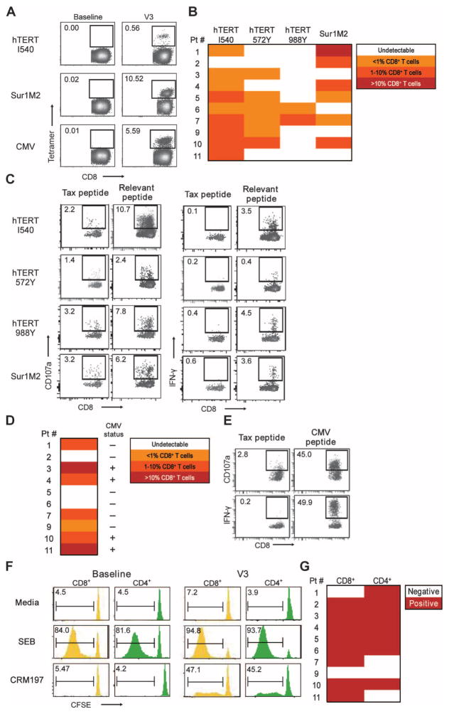
Finally, we studied vaccine-induced immune responses in patients treated with daclizumab and vaccination. We detected robust peptide-specific CD8 T cell responses to tumor vaccine antigens in samples obtained after daclizumab infusion and vaccination (shown for patient 101 in Fig. 4A), with an absence of such responses in all pretreatment control samples. The uniform negativity in these assays at baseline was important because it allowed a straightforward assessment of each patient as either positive or negative, rather than, for example, measuring fold changes from baseline. Peptide-specific responses were observed after as few as two vaccines (Fig. 4A). All 10 evaluable patients responded to at least one tumor peptide (median, two peptides) (Fig. 4B). The vast majority of maximum responses to peptide vaccination occurred at time points of maximal daclizumab-mediated Treg depletion. CD8 T cells induced by vaccination exhibited effector functions, as evidenced by specific mobilization of CD107a and secretion of IFN-γ (Fig. 4C). Responses to CMV peptide were observed before and after vaccination in patients who were CMV-seropositive. In three of six CMV-seronegative patients, responses to CMV peptide were also observed (Fig. 3D), providing evidence for immunological priming in the face of CD25 blockade. CMV-specific T cells observed after vaccination exhibited effector functions (Fig. 4E).
Fig. 4.
Impact of CD25 blockade on vaccine immune responses in patients in vivo. (A to G) Peripheral blood T cells obtained from patients at baseline and at various times after treatment with daclizumab plus an experimental vaccine were analyzed for (A and B) peptide/MHC tetramer reactivity, (C) peptide-specific CD8 T cell responses as measured by CD107a mobilization and IFN-γ expression, (D) CMV tetramer reactivity, (E) CMV-specific CD8 T cell responses as measured by CD107a mobilization and IFN-γ expression, and (F and G) CRM197-specific responses for CD8 and CD4 T cells as measured by antigen-induced CFSE dilution. Representative data for each assay are shown in (A), (C), (E), and (F). Summary data for all patients are shown as heat maps of the best responses in (B), (D), and (G). V3 corresponds to the time of the third vaccine. In (C) and (E), an HLA-A2–binding peptide from HTLV-1 tax (Tax) was used as a negative control, and in (F) and (G), Staphylococcus enterotoxin B (SEB) was used as a positive control. In (D), patient CMV status was determined by seropositivity at baseline; all CMV-seronegative patients remained seronegative at the end of the study.
Patients vigorously responded to CRM197 antigen in response to PCV vaccination. Although no responses were detected at baseline, positive CRM197 responses for CD8 or CD4 T cells were observed for 90% of patients when tested on samples obtained within the window of daclizumab-mediated Treg depletion (median, week 7; range, weeks 2 to 19) (Fig. 4, F and G). Because no patient had been previously treated with CRM197-containing vaccines, these T cell responses likely represent immunological priming to CRM197.
Comparison of vaccination with and without daclizumab
To begin to understand the potential benefit of including daclizumab in cancer vaccination, we compared findings from this current study with those of a previously published study of 19 patients treated with one of the same hTERT peptides but without daclizumab (29). Eligibility criteria for these two studies were identical. The analysis of immune response was based on evaluable patients, defined as patients who received at least four vaccines. Seventeen patients on the previous hTERT peptide study and 10 patients on the hTERT plus daclizumab study were evaluable for immune response assessment. Using identical criteria for assay positivity, we found that the immune response rate to the I540 hTERT peptide on the hTERT plus daclizumab study was higher than the response rate on the previous hTERT-only study. The hTERT I540 immune response rate was 90% (9 of 10 patients) in the hTERT plus daclizumab study (90% exact CI, 60.6 to 99.9%) versus 76.5% (13 of 17 patients) immune response rate in the hTERT-only study (90% CI, 53.9 to 91.5%). Sample sizes in each study were small, having been calculated for the purpose of addressing the primary clinical endpoint of each study, which was safety, rather than the secondary endpoint of immune response rate. Thus, although the immune response rate with daclizumab is higher than with peptide alone, the difference did not reach statistical significance, and a larger study is warranted.
We also explored whether daclizumab interferes with vaccine-induced immune responses, as has been suggested in a previous study (15). Again, comparing our current study to our previously published study of hTERT vaccination without daclizumab, we found that hTERT plus daclizumab was not inferior to hTERT alone with regard to immune response—daclizumab does not interfere with vaccine-induced immune responses. By simulation, we determined that the Bayesian posterior probability, that the true immune response rate of hTERT plus daclizumab is greater than that of hTERT alone, is 0.766. This probability calculation assumed independent uninformative β priors and observed immune responses in 9 of 10 patients on hTERT plus daclizumab and in 13 of 17 patients on hTERT alone.
Finally, patients in the two clinical studies were analyzed for overall survival after an intent-to-treat analysis plan in which all treated patients, regardless of immune response evaluability, are included in the analysis. The median overall survival on the hTERT plus daclizumab study (27.8 months; 95% CI, 18.7 to 36.9 months) was longer than on the hTERT-only study (20.9 months; 95% CI, 12.2 to 29.6 months). Although the difference in overall survival was marked, again limited by sample size, it did not reach statistical significance.
DISCUSSION
It is now well accepted that Tregs cripple productive immune surveillance and are a major obstacle for the development of active immunotherapy for cancer. Here, we show that in vivo CD25 blockade in patients with breast cancer—accomplished safely with a single infusion of the FDA-approved CD25-blocking mAb daclizumab—results in an acute and prolonged depletion of circulating Tregs that is greater and more sustained than other CD25-targeting agents previously reported [summarized in (32)]. For several patients in our study, the systemic burden of both CD25+ FoxP3+ and total FoxP3+ Tregs never recovered to baseline levels and reset within the range for healthy individuals of comparable age. Nevertheless, we show that daclizumab permits robust priming and boosting of effector T cell responses to vaccination in vivo and does so without causing autoimmunity. We conclude that long-term CD25 blockade represents an approach to circumvent a major element of immune suppression in patients with cancer.
We also conclude that IL-2 deprivation at least in large part explains the daclizumab effect on Tregs. In mechanistic in vitro studies, we found that CD25 blockade results in loss of both FoxP3 expression and Treg suppressive function. These changes are consistent with Treg reprogramming that has previously been described in murine models (18, 33). In addition, we were unable to demonstrate a role for daclizumab-mediated antibody-dependent cellular cytotoxicity or complement-mediated cytotoxicity, despite its IgG1 structure, further suggesting that cytokine deprivation and Treg reprogramming are the dominant mechanisms of daclizumab Treg depletion in vivo. We found that blocking IL-2 signaling via daclizumab does not affect all Tregs the same, but rather, it selectively interferes with FoxP3 expression in CD45RAneg Tregs, a subset prominent in cancer patients and likely to be induced by progressively growing cancers. CD45RA+ Tregs, on the other hand, do not appear to be affected by CD25 blockade with regard to FoxP3 expression or function.
The implication of these observations is that CD45RA+ Tregs might serve as a stop gap against systemic autoimmunity that has been observed in mice and humans with global Treg depletion. Indeed, screening for the induction of autoimmunity was the chief objective of our clinical study of daclizumab. In the landmark paper from Sakaguchi and colleagues in 1995 (13), a wide range of autoimmune disorders including gastritis, thyroiditis, glomerulonephritis, and polyarthritis developed in mice after adoptive transfers of CD25-depleted T cells. Moreover, CD25 knockout mice develop autoimmune hemolytic anemia and inflammatory bowel disorders (34), and mice treated with neutralizing anti–IL-2 antibody develop T cell–mediated gastritis, thyroiditis, and neuropathy (5). In our clinical study, however, no such autoimmune disorders were observed in patients after daclizumab despite careful clinical screening. Potentially important in reconciling these observations in mice and humans is the fact that daclizumab is not directly depleting but rather, as we show here, acts on a subset of Tregs via cytokine deprivation. Thus, we hypothesize that after CD25 blockade, CD45RAneg Tregs are reprogrammed but CD45RA+ Tregs are not, and these latter cells may guard against systemic autoimmunity.
Clinically, Treg reprogramming via CD25 blockade is a possible benefit that would nevertheless have to balance with potential impairment of dendritic cell potency and interference with effector responses recently described for the drug in other experimental systems (15, 16, 35). This balance will likely critically depend on daclizumab dose and schedule and, importantly, on the clinical setting as well (36). For reasons that seem linked to NK cell immune suppression of T cells that can be observed clinically after repeated high doses of daclizumab (37), daclizumab can be effective in treating autoimmune conditions such as multiple sclerosis (30, 31). We also observed changes in NK cell subsets, but these changes were modest in comparison to the multiple sclerosis studies, likely because we used a single dose of daclizumab.
Our findings and conclusions are in contrast to one previously published study of daclizumab used in concert with a melanoma vaccine (15). There, the investigators concluded that daclizumab interfered with the induction of functional, tumor-specific effector T cells and antibodies, although vaccine-induced keyhole limpet hemocyanin–specific T cell responses were not adversely affected (15). However, major differences in the clinical design between this study and ours may explain the apparent discrepancies in the two studies. For example, in the melanoma study, the vaccine was composed of antigen-loaded, ex vivo–produced dendritic cells, which are well described to express CD25 and therefore potentially susceptible to daclizumab inhibition (16). In addition, key conclusions of the study were based on analysis of delayed-type hypersensitivity (DTH) responses in a small subset of patients. Of those patients with DTH positivity, functional T cells isolated from the DTH sites were detected in two of two (100%) patients in the control cohort and in one of five (20%) patients in the daclizumab cohort (15). Although these rates appear to be dissimilar, due to the extremely small sample sizes, the precision is low and they are not statistically different (P = 0.14, Fisher’s exact test). In our study, we observed consistent robust CD8 and CD4 T cell priming and boosting to all vaccine antigens in the wake of daclizumab-mediated Treg depletion, suggesting that the clinical setting and the vaccine formulation used are critical variables in this approach that warrant further study.
In summary, we conclude that CD25 blockade depletes and selectively reprograms Tregs in concert with active immune therapy in cancer patients. These results suggest a mechanism to target cancer-associated Tregs while avoiding autoimmunity.
MATERIALS AND METHODS
Purification and isolation of T cells and Tregs
Primary human CD4 T cells were obtained from the University of Pennsylvania Human Immunology Core as de-identified, anonymous samples from healthy donors who gave informed consent to undergo apheresis. These cells were stained with V450-CD4 clone RPA-T4, PE-CD25 clone MA-251, and FITC-CD45RA clone HI100 and sorted into CD4+ CD25neg CD45RA+ (naïve), CD4+ CD25neg CD45RAneg (memory), CD4+ CD25high CD45RA+ (CD45RA+ Treg), and CD4+ CD25high CD45RAneg (CD45RAneg Treg) using an Influx jet-in-air cell sorter with Spigot software (BD Biosciences). In our standard configuration with a 70-μm nozzle, sheath pressure was 35 psi with a drop drive frequency of 79.1 kHz and piezo amplitude of 4.27 V, resulting in a drop delay of 35.9 drops. We used a standard forward scatter threshold and logarithmic amplifiers for all fluorescent parameters. Forward scatter pulse heights versus area parameters were used for aggregate detection. Samples were run at trigger rates of about 12,000 to 18,000 cells/s with efficiencies greater than 90%.
Treg assays
Purified T cell populations were incubated in vitro in the presence of IL-2 (20 U/ml, Novartis) and either daclizumab (10 μg/ml) or human IgG1 (Sigma-Aldrich) (10 μg/ml). To measure Treg viability, cells were mixed with Guava ViaCount reagent (Guava Technologies) for 10 min, and viable cells were then quantified using a Guava Personal Analyzer flow cytometer (Guava Technologies) per the manufacturer’s specifications. Carboxyfluorescein diacetate succinimidyl ester (CFSE)–based CD4 T cell suppression assays to monitor Treg function were performed as previously described (28, 38). Data were acquired on an LSR II flow cytometer using the FACSDiva software analyzed using FlowJo software package. Percent suppression was calculated using the following formula: 1 – number of effector T cell divisions in suppressed condition divided by the number of effector T cell divisions in unsuppressed condition × 100. Assays to measure Treg production of IFN-γ after phorbol 12-myristate 13-acetate (PMA) and ionomycin treatment were performed as previously described (28).
Flow cytometry
Flow cytometry was performed with a FACSCanto cytometer and FACSDiva software (BD Biosciences). Fluorochrome-conjugated mAbs used were allophycocyanin (APC)– and phycoerythrin (PE)–Cy7–CD3 clone SK7, fluorescein isothiocyanate (FITC)–, APC-, and V450-CD4 clone RPA-T4, peridinin chlorophyll protein (PerCP)–CD4 clone SK3, V450-CD8 clone RPA-T8, PerCP-CD14 clone MφP9, FITC-CD16 clone 3G8, PerCP-CD19 clone 4G7, APC-CD19 clone HIB19, PE-CD25 clone 2A3, APC-CD56 clone B159, and FITC-CD107a clone H4A3 (BD Biosciences); APC-CD8 clone B9.11 (Beckman Coulter); Alexa Fluor 488–FoxP3 clone 259D (BioLegend); APC–anti–IFN-γ clone 4S.B3 (eBioscience); and PE-CD25 clone 4E3 (Miltenyi Biotec). Peptide/major histocompatibility complex (MHC) class I tetramer analysis was performed with soluble peptide/HLA-A2 tetramers (Beckman Coulter Immunomics), with the cutoff for a positive response defined as the mean ± 3 SDs for the percentage of tetramer-positive CD8 T cells among peripheral blood mononuclear cells (PBMCs) from a panel of HLA-A2neg healthy volunteers (that is, 0.1% of CD8 T cells) and a panel of HLA-A2+ healthy volunteers (also 0.1% of CD8 T cells), as previously described (29).
T cell assays
In vitro peptide stimulation of PBMCs to assess immune response was performed as previously described (29). For CD107a and IFN-γ analysis, in vitro–stimulated cells were incubated with CD107a mAb and with T2 cells [2:1 ratio; American Type Culture Collection (ATCC)] loaded with peptide (1 μg/ml) and β2-microglobulin (2.5 μg/ml) (Sigma) or with staphylococcal enterotoxin B (1 ng/ml) (EMD Chemicals) with brefelden A added for 4 hours before intracellular staining for IFN-γ as previously described (39). T cell responses to the CRM197 protein were measured by CFSE staining of responder T cells, with cutoff for positivity being defined as 5% CFSEdim CD4+ or CD8+ T cells, as previously described (40).
Cytolysis assays
To assess antibody-dependent cell cytotoxicity, CFSE-labeled lympho-blastoid tumor cells (Daudi, Ramos, and SR) (ATCC) or CFSE-labeled purified CD4 T cells (>85%) were incubated with PBMCs from designated healthy donors (8 to 15% CD56+) at a PBMC/target ratio of 100:1 for 4 hours at 37°C in the presence of daclizumab, IgG1, or rituximab (Genentech/Biogen Idec) (each at 1 μg/ml) in 10% human AB serum (HS), mixed with 10,000 anti-mouse Igκ CompBeads (BD Biosciences) that had been conjugated to V450-mouse IgG1,κ clone MOPC-21 (BD Biosciences). Cells were then stained with the viability marker 7-aminoactinomycin D (7-AAD) (BD Biosciences) and analyzed by flow cytometry. Alive cells (CFSE+ 7-AADneg) were counted relative to the number of beads and quantified as previously described (41). To assess complement-dependent cytotoxicity, targets were incubated with 10% HS or, as a control, 10% heat-inactivated HS for 4 hours at 37°C, mixed with V450 beads, stained with 7-AAD, and analyzed.
Patients and clinical study design
The clinical protocol was an open-label prospective study of patients with metastatic breast cancer refractory to at least one standard therapy for metastatic disease. The protocol was approved by the University of Pennsylvania Institutional Review Board and conducted with FDA approval of an investigator-sponsored investigational new drug (IND) application. Signed, written informed consent was obtained from each patient.
Eligible patients had to be >18 years of age and HLA-A2+ with a baseline Eastern Cooperative Group Clinical performance status of <2. At baseline, patients had to have adequate hematological function (white blood count >3000 cells/mm3, hemoglobulin >10, and platelet count >75,000 cells/mm3), adequate renal function (serum creatinine <1.5 times upper limit of normal), adequate hepatic function (total bilirubin <1.5 times upper limit of normal, aspartate aminotransferase and alanine aminotransferase <2.5 times upper limit of normal), and imaging studies of the brain negative for metastatic disease. Patients were excluded if pregnant or lactating; if HIV-, HTLV-1 (human T cell leukemia virus–1)–, hepatitis B–, or hepatitis C–positive; for active infection or treatment with chemotherapy, radiation therapy, immunotherapy, immunosuppressive drugs, glucocorticoids, hematopoietic growth factors, or other investigational drugs within 30 days of treatment; or for a history of stem cell transplantation, alcohol abuse, or illicit drug use. Concomitant use of chemotherapy, radiation therapy, steroids, immunosuppressive drugs, anticoagulants, or other investigational drugs was not allowed. Eligibility criteria were identical to those of a previous clinical trial from our group (29).
Patients received a single intravenous infusion of daclizumab (Roche) at 1 mg/kg followed 1 week later by vaccination with five HLA-A2–binding peptides (100 μg/ml per peptide per injection) every other week four times and then monthly until toxicity or clinically important disease progression. Vaccinations consisted of aqueous solutions of hTERT peptides I540 (ILAKFLHWL) (42), 572Y (YLFFYRKSU) (43), 988Y (YLQVNSLQTV) (43), Sur1M2 (LMLGEFLKL) (44), and N495 (NLVPMVATV) from CMV pp65 (45) (each peptide >92% pure and Good Manufacturing Practice–grade; Merck Biosciences AG) emulsified in the adjuvant Montanide ISA 51 VG (Seppic) and delivered subcutaneously in the thighs. Sargramostim (clinical-grade granulocyte-macrophage colony-stimulating factor; Berlex Laboratories) was given subcutaneously at the two injection sites (70 μg per site) with each peptide vaccination. Prevnar (PCV; Wyeth) (0.5 ml per injection) was given intramuscularly in the deltoid at the time of the first, third, and fifth vaccine.
Toxicity was graded according to the National Cancer Institute Common Toxicity Criteria (version 3.0). Tumor response was assessed every 2 to 3 months according to RECIST criteria (version 1.0). Patients receiving fewer than four vaccines were considered unevaluable for clinical response or vaccine-specific immune response and were replaced. Overall survival was defined from the date of first treatment to death or last patient contact. Progression-free survival was defined from the date of first treatment to the first documented disease progression, death, or last patient contact. PBMCs were obtained by phlebotomy of patients on the current study who provided written, informed consent using an Institutional Review Board–approved protocol via Ficoll centrifugation and then frozen at −150°C before use. In some experiments, PBMCs were from patients on a previous peptide vaccine trial for which signed, written informed consent had been obtained (29).
Statistical analysis
Student’s t tests were used to compare means between two independent groups, whereas paired Student’s t tests were used to compare outcomes before and after vaccination within patients. To analyze trends over time in immunologic variables, we used linear random effects models. STATA procedure xtmixed was used to analyze change from baseline. As in repeated-measures analysis of variance (ANOVA), time was defined as a class variable. Natural log transformation was applied to all variables before modeling. A sensitivity analysis (that is, Poisson regression, which allows zero values) was performed and yielded qualitatively similar results. Kaplan-Meier analysis was used to estimate time-to-event outcomes. Analyses were performed with STATA v11 (StataCorp), SPSS v17 (SPSS), or Graph-Pad Prism (GraphPad Software). P < 0.05 was considered statistically significant.
Supplementary Material
Fig. S1. Daclizumab does not elicit cytotoxicity of CD25-expressing (A) lymphoid cell lines or (B) CD4 T cells.
Fig. S2. Clinical trial schema.
Fig. S3. CD45RA and CD45RO phenotype of FoxP3+ CD4 T cells from patients with metastatic breast cancer.
Fig. S4. Daclizumab does not block binding of the CD25 mAb clone 4E3, but daclizumab does block binding of CD25 mAb clone 2A3.
Fig. S5. Change in T cell populations shown separately for each patient.
Fig. S6. Impact of daclizumab on (A) FoxP3neg CD4 T cells and (B) CD8 T cells.
Fig. S7. Impact of daclizumab on natural killer (NK) cell populations.
Table S1. Patient characteristics.
Table S2. Treatment-emergent patient adverse events and abnormal laboratory values.
Table S3. T cell subset analysis after daclizumab (corresponds to Fig. 3B).
Acknowledgments
Funding: Supported by NIH grant CA111377 (to R.H.V.), the Juvenile Diabetes Research Foundation (JDRF) Collaborative Centers for Cell Therapy and the JDRF Center on Cord Blood Therapies for Type 1 Diabetes (to J.L.R.), the Breast Cancer Research Foundation (to R.H.V. and S.M.D.), and the Abramson Family Cancer Research Institute (to R.H.V.).
Footnotes
Author contributions: A.J.R., S.M., N.A.A., D.J.P., C.H.P., J.L.R., and R.H.V. designed the experiments. A.J.R., S.M., N.A.A., T.A.C., J.A.T., and L.I.L. performed the experiments. R.M. was the lead biostatistician for the study. A.J.R., R.M., S.M., N.A.A., D.J.P., K.R.F., S.M.D., J.L.R., and R.H.V. interpreted the data. A.R., C.K.T., A.D., and K.R.F. treated and assessed patients. K.R.F. was the principal investigator of the protocol. R.H.V., R.M., A.R., K.R.F., and S.M.D. wrote the clinical protocol. R.H.V. was IND sponsor. The manuscript was written by R.H.V., edited by A.J.R., R.M., S.M., D.J.P., K.R.F., S.M.D., and J.L.R., and approved by all authors.
Competing interests: R.H.V. and S.M.D. declare a potential financial conflict of interest related to inventorship on a patent regarding hTERT as a tumor-associated antigen for cancer immunotherapy (Cancer immunotherapy and diagnosis using universal tumor associated antigens, including hTERT, U.S. Patent 7851591). The other authors declare that they do not have any competing interests.
REFERENCES AND NOTES
- 1.Woo EY, Chu CS, Goletz TJ, Schlienger K, Yeh H, Coukos G, Rubin SC, Kaiser LR, June CH. Regulatory CD4+CD25+ T cells in tumors from patients with early-stage non-small cell lung cancer and late-stage ovarian cancer. Cancer Res. 2001;61:4766–4772. [PubMed] [Google Scholar]
- 2.Clark CE, Hingorani SR, Mick R, Combs C, Tuveson DA, Vonderheide RH. Dynamics of the immune reaction to pancreatic cancer from inception to invasion. Cancer Res. 2007;67:9518–9527. doi: 10.1158/0008-5472.CAN-07-0175. [DOI] [PubMed] [Google Scholar]
- 3.Sakaguchi S, Yamaguchi T, Nomura T, Ono M. Regulatory T cells and immune tolerance. Cell. 2008;133:775–787. doi: 10.1016/j.cell.2008.05.009. [DOI] [PubMed] [Google Scholar]
- 4.Shevach EM. Mechanisms of Foxp3+ T regulatory cell-mediated suppression. Immunity. 2009;30:636–645. doi: 10.1016/j.immuni.2009.04.010. [DOI] [PubMed] [Google Scholar]
- 5.Setoguchi R, Hori S, Takahashi T, Sakaguchi S. Homeostatic maintenance of natural Foxp3+ CD25+ CD4+ regulatory T cells by interleukin (IL)-2 and induction of autoimmune disease by IL-2 neutralization. J Exp Med. 2005;201:723–735. doi: 10.1084/jem.20041982. [DOI] [PMC free article] [PubMed] [Google Scholar]
- 6.Bayer AL, Yu A, Adeegbe D, Malek TR. Essential role for interleukin-2 for CD4+CD25+ T regulatory cell development during the neonatal period. J Exp Med. 2005;201:769–777. doi: 10.1084/jem.20041179. [DOI] [PMC free article] [PubMed] [Google Scholar]
- 7.Fontenot JD, Rasmussen JP, Gavin MA, Rudensky AY. A function for interleukin 2 in Foxp3-expressing regulatory T cells. Nat Immunol. 2005;6:1142–1151. doi: 10.1038/ni1263. [DOI] [PubMed] [Google Scholar]
- 8.Almeida AR, Legrand N, Papiernik M, Freitas AA. Homeostasis of peripheral CD4+ T cells: IL-2Rα and IL-2 shape a population of regulatory cells that controls CD4+ T cell numbers. J Immunol. 2002;169:4850–4860. doi: 10.4049/jimmunol.169.9.4850. [DOI] [PubMed] [Google Scholar]
- 9.Malek TR, Yu A, Vincek V, Scibelli P, Kong L. CD4 regulatory T cells prevent lethal auto-immunity in IL-2Rβ-deficient mice. Implications for the nonredundant function of IL-2. Immunity. 2002;17:167–178. doi: 10.1016/s1074-7613(02)00367-9. [DOI] [PubMed] [Google Scholar]
- 10.Onizuka S, Tawara I, Shimizu J, Sakaguchi S, Fujita T, Nakayama E. Tumor rejection by in vivo administration of anti-CD25 (interleukin-2 receptor α) monoclonal antibody. Cancer Res. 1999;59:3128–3133. [PubMed] [Google Scholar]
- 11.Shimizu J, Yamazaki S, Sakaguchi S. Induction of tumor immunity by removing CD25+CD4+ T cells: A common basis between tumor immunity and autoimmunity. J Immunol. 1999;163:5211–5218. [PubMed] [Google Scholar]
- 12.Sutmuller RP, van Duivenvoorde LM, van Elsas A, Schumacher TN, Wildenberg ME, Allison JP, Toes RE, Offringa R, Melief CJ. Synergism of cytotoxic T lymphocyte–associated antigen 4 blockade and depletion of CD25+ regulatory T cells in antitumor therapy reveals alternative pathways for suppression of autoreactive cytotoxic T lymphocyte responses. J Exp Med. 2001;194:823–832. doi: 10.1084/jem.194.6.823. [DOI] [PMC free article] [PubMed] [Google Scholar]
- 13.Sakaguchi S, Sakaguchi N, Asano M, Itoh M, Toda M. Immunologic self-tolerance maintained by activated T cells expressing IL-2 receptor alpha-chains (CD25). Breakdown of a single mechanism of self-tolerance causes various autoimmune diseases. J Immunol. 1995;155:1151–1164. [PubMed] [Google Scholar]
- 14.Taguchi O, Takahashi T. Administration of anti-interleukin-2 receptor α antibody in vivo induces localized autoimmune disease. Eur J Immunol. 1996;26:1608–1612. doi: 10.1002/eji.1830260730. [DOI] [PubMed] [Google Scholar]
- 15.Jacobs JF, Punt CJ, Lesterhuis WJ, Sutmuller RP, Brouwer HM, Scharenborg NM, Klasen IS, Hilbrands LB, Figdor CG, de Vries IJ, Adema GJ. Dendritic cell vaccination in combination with anti-CD25 monoclonal antibody treatment: A phase I/II study in metastatic melanoma patients. Clin Cancer Res. 2010;16:5067–5078. doi: 10.1158/1078-0432.CCR-10-1757. [DOI] [PubMed] [Google Scholar]
- 16.Wuest SC, Edwan JH, Martin JF, Han S, Perry JS, Cartagena CM, Matsuura E, Maric D, Waldmann TA, Bielekova B. A role for interleukin-2 trans-presentation in dendritic cell–mediated T cell activation in humans, as revealed by daclizumab therapy. Nat Med. 2011;17:604–609. doi: 10.1038/nm.2365. [DOI] [PMC free article] [PubMed] [Google Scholar]
- 17.Malek TR. The biology of interleukin-2. Annu Rev Immunol. 2008;26:453–479. doi: 10.1146/annurev.immunol.26.021607.090357. [DOI] [PubMed] [Google Scholar]
- 18.Zhou X, Bailey-Bucktrout S, Jeker LT, Bluestone JA. Plasticity of CD4+ FoxP3+ T cells. Curr Opin Immunol. 2009;21:281–285. doi: 10.1016/j.coi.2009.05.007. [DOI] [PMC free article] [PubMed] [Google Scholar]
- 19.Rubtsov YP, Niec RE, Josefowicz S, Li L, Darce J, Mathis D, Benoist C, Rudensky AY. Stability of the regulatory T cell lineage in vivo. Science. 2010;329:1667–1671. doi: 10.1126/science.1191996. [DOI] [PMC free article] [PubMed] [Google Scholar]
- 20.Leonard WJ, Depper JM, Uchiyama T, Smith KA, Waldmann TA, Greene WC. A monoclonal antibody that appears to recognize the receptor for human T-cell growth factor; partial characterization of the receptor. Nature. 1982;300:267–269. doi: 10.1038/300267a0. [DOI] [PubMed] [Google Scholar]
- 21.Rech AJ, Vonderheide RH. Clinical use of anti-CD25 antibody daclizumab to enhance immune responses to tumor antigen vaccination by targeting regulatory T cells. Ann N Y Acad Sci. 2009;1174:99–106. doi: 10.1111/j.1749-6632.2009.04939.x. [DOI] [PubMed] [Google Scholar]
- 22.Mitchell DA, Cui X, Schmittling RJ, Sanchez-Perez L, Snyder DJ, Congdon KL, Archer GE, Desjardins A, Friedman AH, Friedman HS, Herndon JE, II, McLendon RE, Reardon DA, Vredenburgh JJ, Bigner DD, Sampson JH. Monoclonal antibody blockade of IL-2 receptor α during lymphopenia selectively depletes regulatory T cells in mice and humans. Blood. 2011;118:3003–3012. doi: 10.1182/blood-2011-02-334565. [DOI] [PMC free article] [PubMed] [Google Scholar]
- 23.van de Linde P, Tysma OM, Medema JP, Hale G, Waldmann H, Roelen DL, Roep BO. Mechanisms of antibody immunotherapy on clonal islet reactive T cells. Hum Immunol. 2006;67:264–273. doi: 10.1016/j.humimm.2006.02.027. [DOI] [PubMed] [Google Scholar]
- 24.Hoffmann P, Eder R, Boeld TJ, Doser K, Piseshka B, Andreesen R, Edinger M. Only the CD45RA+ subpopulation of CD4+CD25high T cells gives rise to homogeneous regulatory T-cell lines upon in vitro expansion. Blood. 2006;108:4260–4267. doi: 10.1182/blood-2006-06-027409. [DOI] [PubMed] [Google Scholar]
- 25.Miyara M, Yoshioka Y, Kitoh A, Shima T, Wing K, Niwa A, Parizot C, Taflin C, Heike T, Valeyre D, Mathian A, Nakahata T, Yamaguchi T, Nomura T, Ono M, Amoura Z, Gorochov G, Sakaguchi S. Functional delineation and differentiation dynamics of human CD4+ T cells expressing the FoxP3 transcription factor. Immunity. 2009;30:899–911. doi: 10.1016/j.immuni.2009.03.019. [DOI] [PubMed] [Google Scholar]
- 26.Golovina TN, Mikheeva T, Brusko TM, Blazar BR, Bluestone JA, Riley JL. Retinoic acid and rapamycin differentially affect and synergistically promote the ex vivo expansion of natural human T regulatory cells. PLoS One. 2011;6:e15868. doi: 10.1371/journal.pone.0015868. [DOI] [PMC free article] [PubMed] [Google Scholar]
- 27.Bates GJ, Fox SB, Han C, Leek RD, Garcia JF, Harris AL, Banham AH. Quantification of regulatory T cells enables the identification of high-risk breast cancer patients and those at risk of late relapse. J Clin Oncol. 2006;24:5373–5380. doi: 10.1200/JCO.2006.05.9584. [DOI] [PubMed] [Google Scholar]
- 28.Rech AJ, Mick R, Kaplan DE, Chang KM, Domchek SM, Vonderheide RH. Homeostasis of peripheral FoxP3+ CD4+ regulatory T cells in patients with early and late stage breast cancer. Cancer Immunol Immunother. 2010;59:599–607. doi: 10.1007/s00262-009-0780-x. [DOI] [PMC free article] [PubMed] [Google Scholar]
- 29.Domchek SM, Recio A, Mick R, Clark CE, Carpenter EL, Fox KR, DeMichele A, Schuchter LM, Leibowitz MS, Wexler MH, Vance BA, Beatty GL, Veloso E, Feldman MD, Vonderheide RH. Telomerase-specific T-cell immunity in breast cancer: Effect of vaccination on tumor immunosurveillance. Cancer Res. 2007;67:10546–10555. doi: 10.1158/0008-5472.CAN-07-2765. [DOI] [PubMed] [Google Scholar]
- 30.Bielekova B, Richert N, Howard T, Blevins G, Markovic-Plese S, McCartin J, Frank JA, Würfel J, Ohayon J, Waldmann TA, McFarland HF, Martin R. Humanized anti-CD25 (daclizumab) inhibits disease activity in multiple sclerosis patients failing to respond to interferon β. Proc Natl Acad Sci USA. 2004;101:8705–8708. doi: 10.1073/pnas.0402653101. [DOI] [PMC free article] [PubMed] [Google Scholar]
- 31.Wynn D, Kaufman M, Montalban X, Vollmer T, Simon J, Elkins J, O’Neill G, Neyer L, Sheridan J, Wang C, Fong A, Rose JW. CHOICE investigators, Daclizumab in active relapsing multiple sclerosis (CHOICE study): A phase 2, randomised, double-blind, placebo-controlled, add-on trial with interferon beta. Lancet Neurol. 2010;9:381–390. doi: 10.1016/S1474-4422(10)70033-8. [DOI] [PubMed] [Google Scholar]
- 32.Golovina TN, Vonderheide RH. Regulatory T cells: Overcoming suppression of T-cell immunity. Cancer J. 2010;16:342–347. doi: 10.1097/PPO.0b013e3181eb336d. [DOI] [PubMed] [Google Scholar]
- 33.Oldenhove G, Bouladoux N, Wohlfert EA, Hall JA, Chou D, Dos Santos L, O’Brien S, Blank R, Lamb E, Natarajan S, Kastenmayer R, Hunter C, Grigg ME, Belkaid Y. Decrease of Foxp3+ Treg cell number and acquisition of effector cell phenotype during lethal infection. Immunity. 2009;31:772–786. doi: 10.1016/j.immuni.2009.10.001. [DOI] [PMC free article] [PubMed] [Google Scholar]
- 34.Willerford DM, Chen J, Ferry JA, Davidson L, Ma A, Alt FW. Interleukin-2 receptor α chain regulates the size and content of the peripheral lymphoid compartment. Immunity. 1995;3:521–530. doi: 10.1016/1074-7613(95)90180-9. [DOI] [PubMed] [Google Scholar]
- 35.Mnasria K, Lagaraine C, Velge-Roussel F, Oueslati R, Lebranchu Y, Baron C. Anti-CD25 antibodies affect cytokine synthesis pattern of human dendritic cells and decrease their ability to prime allogeneic CD4+ T cells. J Leukoc Biol. 2008;84:460–467. doi: 10.1189/jlb.1007712. [DOI] [PubMed] [Google Scholar]
- 36.Schluns KS. Window of opportunity for daclizumab. Nat Med. 2011;17:545–547. doi: 10.1038/nm0511-545. [DOI] [PubMed] [Google Scholar]
- 37.Bielekova B, Catalfamo M, Reichert-Scrivner S, Packer A, Cerna M, Waldmann TA, McFarland H, Henkart PA, Martin R. Regulatory CD56bright natural killer cells mediate immunomodulatory effects of IL-2Rα-targeted therapy (daclizumab) in multiple sclerosis. Proc Natl Acad Sci USA. 2006;103:5941–5946. doi: 10.1073/pnas.0601335103. [DOI] [PMC free article] [PubMed] [Google Scholar]
- 38.Golovina TN, Mikheeva T, Suhoski MM, Aqui NA, Tai VC, Shan X, Liu R, Balcarcel RR, Fisher N, Levine BL, Carroll RG, Warner N, Blazar BR, June CH, Riley JL. CD28 costimulation is essential for human T regulatory expansion and function. J Immunol. 2008;181:2855–2868. doi: 10.4049/jimmunol.181.4.2855. [DOI] [PMC free article] [PubMed] [Google Scholar]
- 39.Carpenter EL, Vance BA, Klein RS, Voloschin A, Dalmau J, Vonderheide RH. Functional analysis of CD8+ T cell responses to the onconeural self protein cdr2 in patients with paraneoplastic cerebellar degeneration. J Neuroimmunol. 2008;193:173–182. doi: 10.1016/j.jneuroim.2007.10.014. [DOI] [PubMed] [Google Scholar]
- 40.Rapoport AP, Stadtmauer EA, Aqui N, Badros A, Cotte J, Chrisley L, Veloso E, Zheng Z, Westphal S, Mair R, Chi N, Ratterree B, Pochran MF, Natt S, Hinkle J, Sickles C, Sohal A, Ruehle K, Lynch C, Zhang L, Porter DL, Luger S, Guo C, Fang HB, Blackwelder W, Hankey K, Mann D, Edelman R, Frasch C, Levine BL, Cross A, June CH. Restoration of immunity in lymphopenic individuals with cancer by vaccination and adoptive T-cell transfer. Nat Med. 2005;11:1230–1237. doi: 10.1038/nm1310. [DOI] [PubMed] [Google Scholar]
- 41.Cao LF, Krymskaya L, Tran V, Mi S, Jensen MC, Blanchard S, Kalos M. Development and application of a multiplexable flow cytometry-based assay to quantify cell-mediated cytolysis. Cytometry A. 2010;77:534–545. doi: 10.1002/cyto.a.20887. [DOI] [PubMed] [Google Scholar]
- 42.Vonderheide RH, Hahn WC, Schultze JL, Nadler LM. The telomerase catalytic subunit is a widely expressed tumor-associated antigen recognized by cytotoxic T lymphocytes. Immunity. 1999;10:673–679. doi: 10.1016/s1074-7613(00)80066-7. [DOI] [PubMed] [Google Scholar]
- 43.Gross DA, Graff-Dubois S, Opolon P, Cornet S, Alves P, Bennaceur-Griscelli A, Faure O, Guillaume P, Firat H, Chouaib S, Lemonnier FA, Davoust J, Miconnet I, Vonderheide RH, Kosmatopoulos K. High vaccination efficiency of low-affinity epitopes in antitumor immunotherapy. J Clin Invest. 2004;113:425–433. doi: 10.1172/JCI19418. [DOI] [PMC free article] [PubMed] [Google Scholar]
- 44.Andersen MH, Pedersen LO, Becker JC, Straten PT. Identification of a cytotoxic T lymphocyte response to the apoptosis inhibitor protein survivin in cancer patients. Cancer Res. 2001;61:869–872. [PubMed] [Google Scholar]
- 45.McLaughlin-Taylor E, Pande H, Forman SJ, Tanamachi B, Li CR, Zaia JA, Greenberg PD, Riddell SR. Identification of the major late human cytomegalovirus matrix protein pp65 as a target antigen for CD8+ virus-specific cytotoxic T lymphocytes. J Med Virol. 1994;43:103–110. doi: 10.1002/jmv.1890430119. [DOI] [PubMed] [Google Scholar]
Associated Data
This section collects any data citations, data availability statements, or supplementary materials included in this article.
Supplementary Materials
Fig. S1. Daclizumab does not elicit cytotoxicity of CD25-expressing (A) lymphoid cell lines or (B) CD4 T cells.
Fig. S2. Clinical trial schema.
Fig. S3. CD45RA and CD45RO phenotype of FoxP3+ CD4 T cells from patients with metastatic breast cancer.
Fig. S4. Daclizumab does not block binding of the CD25 mAb clone 4E3, but daclizumab does block binding of CD25 mAb clone 2A3.
Fig. S5. Change in T cell populations shown separately for each patient.
Fig. S6. Impact of daclizumab on (A) FoxP3neg CD4 T cells and (B) CD8 T cells.
Fig. S7. Impact of daclizumab on natural killer (NK) cell populations.
Table S1. Patient characteristics.
Table S2. Treatment-emergent patient adverse events and abnormal laboratory values.
Table S3. T cell subset analysis after daclizumab (corresponds to Fig. 3B).


