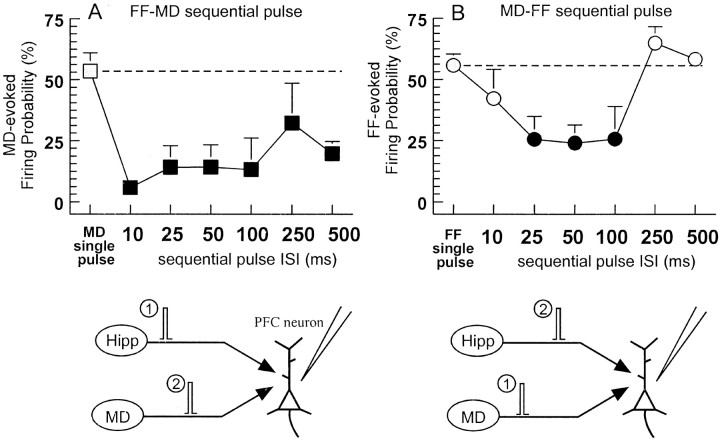
Fig. 2.
Examples of sequential-pulse interactions of the PFC and MD inputs to the PFC. A, In neurons that fired in response to stimulation of both the FF and MD, a conditioning pulse applied to the FF inhibited firing evoked by a test pulse to the MD (black squares; mean + SEM) compared with a single MD pulse alone with the same intensity (gray square, hatched line). B, In these same neurons, a conditioning pulse applied to the MD significantly inhibited firing evoked by an FF test pulse compared with firing evoked by single pulses to the FF (gray circles). Here, the inhibition was significant (p < 0.05) only at intervals of 25–100 msec (black circles) but not at intervals of 10 or 250–500 msec (white circles) when compared with the firing probability observed using single-pulse stimulation (gray circle). Bottom panels diagram the stimulation protocols used forA and B, respectively.
